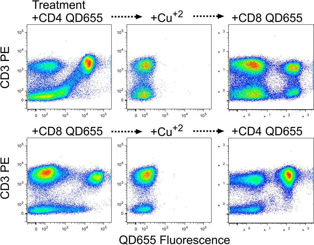
Figure 4. Potential utility of the elimination of fluorescence.
PBMC were stained with PE-anti-CD3 and QD655 conjugated to either anti-CD4 (top) or anti-CD8 (bottom). After analysis (left), cells were treated for 30 min with 30 µM cupric sulfate and reanalyzed (middle). Finally, cells were incubated for 30 min with 1 mM EDTA and washed three times, then re-stained with QD655 conjugated to either anti-CD8 (top) or anti-CD4 (bottom).

