
Fig. 6.
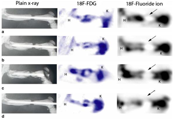
Plain AP radiograph and corresponding 18F-FDG and 18F-fluoride PET scan images of a femur from group II at 1 week (a), 2 weeks (b), 3 weeks (c), and 4 weeks (d) after surgery. Plain radiographs reveal the maintenance of a bony gap and no bridging bone at the surgical site even 4 weeks after surgery (d). Corresponding PET images using 18F-fluoride show tracer uptake extending into the fracture site from the bony ends; however, signal intensity is minimal at all time points, revealing poor biologic healing potential. Despite the absence of bone formation within the fracture site, significant localized uptake of 18F-FDG is seen at all time points. K knee, arrows nonunion site, H hip