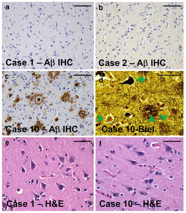
Fig. 1.

Representative photomicrographs from the superior and middle temporal cortical gyri of individuals sampled for the current study. Tissue sections for histopathology (Table 1) were immediately adjacent to the tissue samples used for RNA isolation. Amyloid plaques are stained using Aβ immunohistochemistry (IHC) as shown in a–c. Note that in cases 1 and 2 (a, b), there is no observable Aβ plaques. By contrast, case 10 (c, d, f) had the highest densities of Alzheimer's disease (AD)-related pathology. Bielschowsky (Biel) silver stain (d) confirms the presence in case 10 of diffuse amyloid plaques (downward arrow), neuritic amyloid plaques (upward arrow), neurofibrillary tangles (leftward arrow), and also viable-appearing neurons (rightward arrow). Hematoxylin and eosin (H&E) stains of both cases 1 and 10 demonstrate viable-appearing pyramidal neurons in the temporal cortex. Even in the most severely affected AD brain evaluated in the present study, the disease had not progressed to a stage of widespread neuronal cell death. Scale bars 100 μm (a–c) and 50 μm (d–f)
