
Fig. 2.
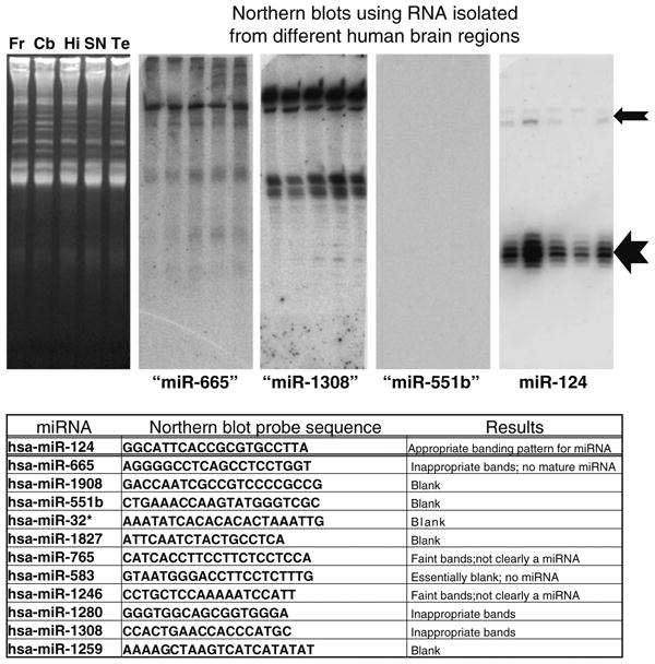
Northern blots were performed on ten different miRNAs with relatively high expression according to the microarrays used in the current studies despite lack of previous evidence of brain expression. 20 μg of RNA was isolated from human frontal cortex (Fr), cerebellum (Cb), hippocampus CA1 (Hi), substantia nigra (SN), and superior and middle temporal gyri (Te), and these samples were run on 15% urea-polyacrylamide gel electrophoresis. Representative ethidium-bromide stained gel is shown in left. Results are shown for miR-665, miR-1308, mir-551b, and miR-124 with the latter being the only one that has been firmly documented in human brain. Note that the northern blotting staining pattern for miR-124 is exactly as expected with a ∼22 nts band (large arrow) and a slower-migrating ∼70 nts band (smaller arrow). However, none of the other putative miRNAs stained as expected for a conventional miRNA (see table). These results, along with prior studies, confirm that some annotated miRNAs are not necessarily functioning as conventional miRNAs in human brain