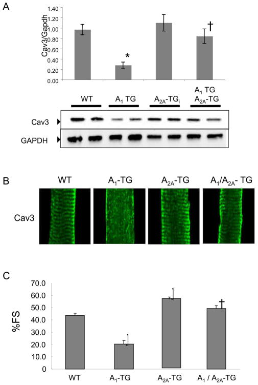
Figure 5.
Caveolin (Cav-3) protein expression, caveolin-3 localization and cardiac function in transgenic mice overexpressing both A1-R and A2A-R. (A) caveolin-3 expression in WT, A1-TG, A2A-TG and A1/A2A-TG mice. Ventricular extracts from 10-week-old age and sex matched mice (N=4) were probed with indicated antibodies. Signals were normalized to GAPDH expression in WT hearts and were analyzed by non-parametric method. Analyzed values were mean+/-SE, *P<0.05 vs WT. (B) Caveolin-3 localization in cardiac myocytes. 600X confocal images are shown. (C) Percent fractional shortening (%FS) of indicated mouse groups. *P<0.001 vs WT, †P<0.001 vs A1TG. (n=15–21, SE, male 8–12 week old mice).

