
Figure 8.
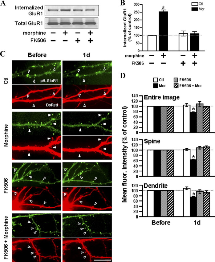
Inhibition of calcineurin blocks the morphine-regulated trafficking of GluR1. A, Surface biotinylated hippocampal neurons were incubated in the absence (left) or presence (right) of 1 μm FK506 for 45 min, followed by treatment with morphine (10 μm; 1 d). Top, The internalized GluR1 subunits were precipitated with immobilized streptavidin and detected with an anti-GluR1 antibody by Western blotting. Bottom, Total GluR1 subunits from whole-cell lysates were detected for a comparison. B, Densitometric quantifications of Western blots in A on the internalized GluR1 (normalized to the control without any drug treatment; p < 0.05; n = 3). C, Neurons coexpressing pHluorin-GluR1 and DsRed were imaged before (left) and 1 d (right) after treatment with no drug, 10 μm morphine, 1 μm FK506, or FK506 with morphine. Open arrowheads denote no change in pHluorin-GluR1 fluorescence and spine morphology. Solid arrowheads denote a decrease in the pHluorin-GluR1 fluorescence and spine morphology by morphine. Scale bar, 10 μm. D, Mean fluorescence (fluor.) intensities of pHluorin-GluR1 from different regions of neurons (the entire image, spines, and dendritic shafts) were normalized to data before treatments in the four groups of experiments [no drug (Ctl), morphine (Mor), FK506, or FK506 with morphine]. *p < 0.05, compared with data before treatment. At least five dendrites of single neurons were analyzed (n = 15 in each group). Error bars represent ± SEM.