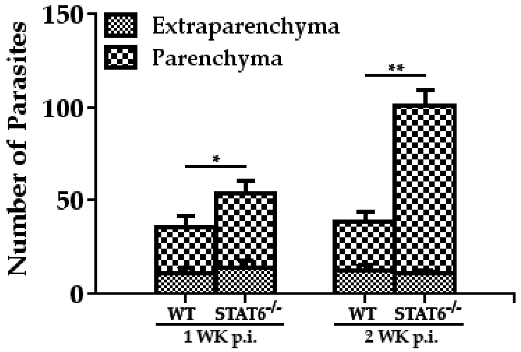
Figure 6. CNS infection with M. corti in STAT6−/− mice resulted in increased parasite burden in CNS.
The WT (C57BL/6) and STAT6−/− were i.c. infected with ~40 M. corti metacestodes. After 1 wk and 2 wk of infection, mice were sacrificed and the location and number of parasites was enumerated in parenchymal and extraparenchymal regions by microscopic examination of serial H&E-stained brain sections. The significant differences in parasite numbers in M. corti infected STAT6−/− as compared with the infected WT animals are denoted by asterisks (*, p<0.05; **, p<0.005). Results are from three independent experiments.

