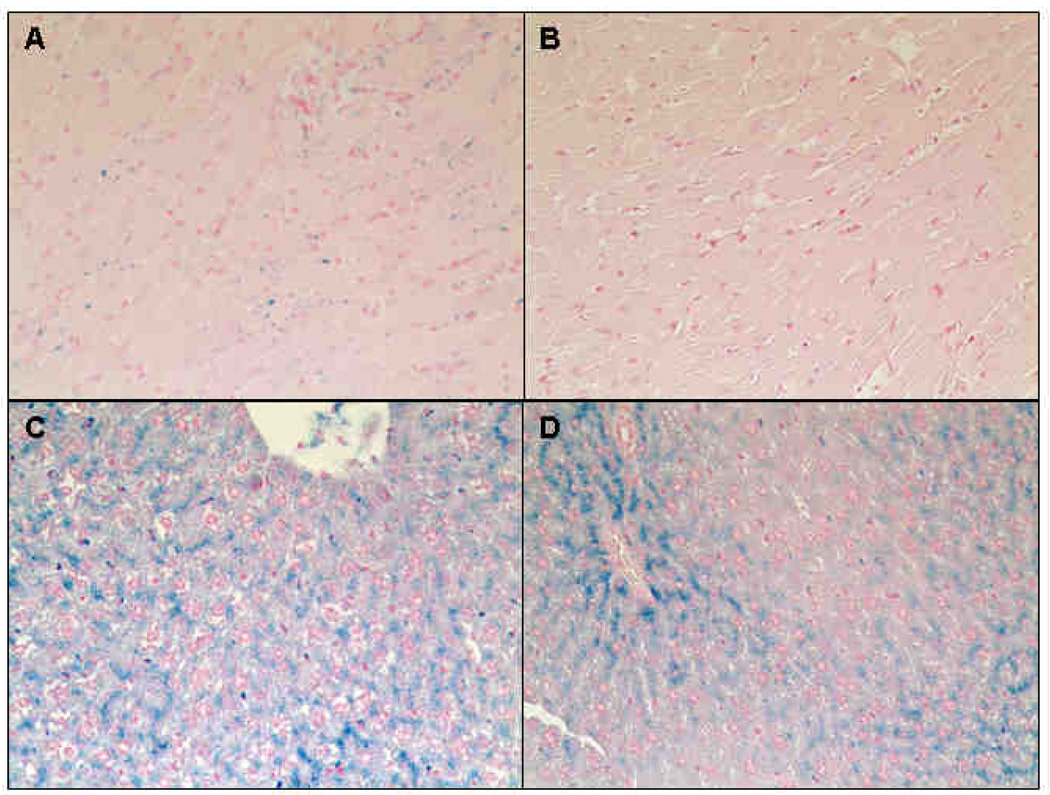
Figure 6. Representative histological examination of heart and liver following verapamil treatment.
Specimens were stained with Perl’s Prussian blue iron stain (A-D). Cardiac sections from an untreated animal is shown in Figure 6A. Iron was predominantly found in cardiomyocytes. Only minor iron staining was detectable in control animals treated with verapamil (B). In untreated animals, hepatic iron mainly accumulated in parenchyma cells, but not in Kuffper cells (C). Verapamil treatment cleared hepatocytes (D). Original magnifications is x200.

