Figure 1.
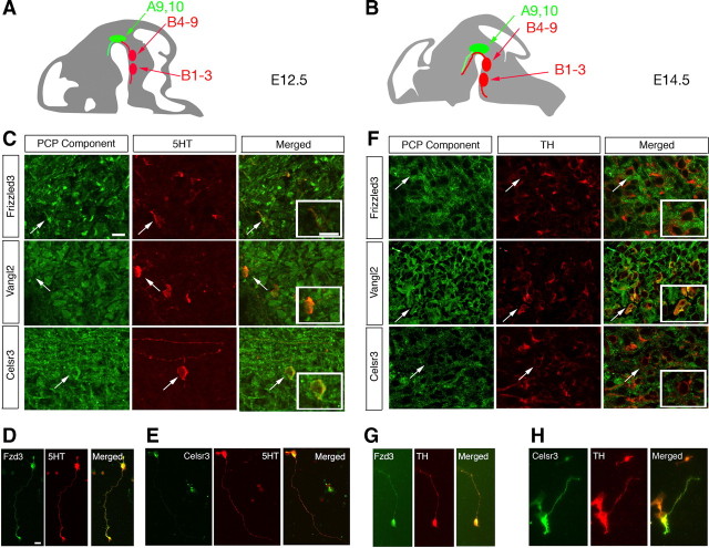
PCP signaling components are expressed in 5-HT- and TH-positive neurons during midbrain and hindbrain development. Schematics in A and B indicate the anatomical location of the descending and ascending serotonin systems (B1–B3 and B4–B9; red) and the mesodiencephalic dopamine system (A9, A10; green) in mouse at E12.5 and E14.5, respectively. C, Immunohistochemistry for 5-HT in red and PCP receptors in green in sagittal sections through the embryonic hindbrain at E12.5. Frizzled3, Vangl2, and Celsr3 all colocalize with 5-HT-positive neurons and fibers. Insets depict higher-magnification images of cells indicated by the arrow. D, Confocal images showing immunohistochemistry for TH in red and PCP receptors in green in coronal sections through the E14.5 midbrain. Frizzled3, Vangl2, and Celsr3 all colocalize with TH-positive neurons and fibers. Insets depict higher-magnification images of cells indicated by the arrow. D, E, 5-HT-positive dissociated neurons coimmunostained with 5-HT in red and Fzd3 (D) or Celsr3 (E) in green. G, H, TH-positive dissociated neurons coimmunostained with TH in red and Fzd3 (G) or Celsr3 (H) in green. Scale bars, 20 μm.

