Figure 3.
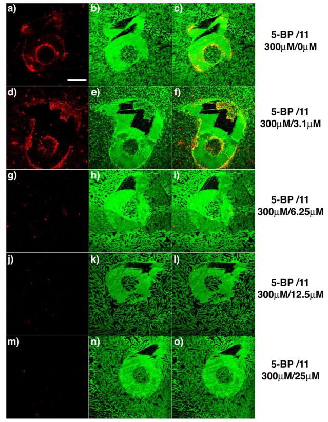
Titration of 11 against active TG2 in a fibroblast scratch assay. TG2 activity was visualized in situ after scratching a confluent WI-38 monolayer with a small pipette tip. Significant TG2 activity was detected around the wound in the presence of 300 μM 5BP and vehicle (DMSO) (a-c). Clear inhibition of active TG2 was observed in the presence of 6.25 μM 11 (g-i), and complete inhibition was observed at or above 12.5 μM 11 (j-o). In this assay, active TG2 was detected by exposing fixed cultures to streptavidin Alexa fluor 555 (a, d, g, j, m). The scratch geometry is conveniently visualized by co-staining with polyclonal anti-fibronectin antibody, followed by a secondary antibody conjugated Alexa fluor 488 (b, e, h, k, n). Overlays of the left and middle images are in the right column (c, f, i, l, o). Scale bar represents 200 μm and applies to all panels.

