Figure 1.
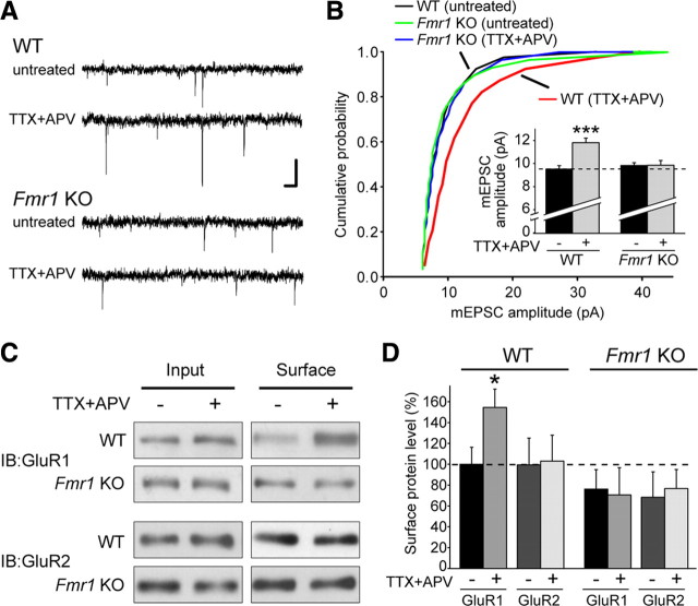
FMRP is required for TTX + APV-induced synaptic scaling. A, Representative mEPSC traces from wild-type and Fmr1 knock-out (untreated and TTX + APV treated) neurons in hippocampal slice culture. Calibration: 10 pA, 40 ms. B, Cumulative distribution of mEPSC amplitudes from WT and KO neurons treated with 36 h of TTX + APV (p < 0.001, Kolmogorov–Smirnov test). Inset, Quantification of average mEPSC amplitude (n = 28–34; ***p < 0.001). C, Representative blots for biotinylation of surface AMPARs in primary cultured neurons after 24 h of TTX + APV treatment. IB, Immunoblot. D, Quantification of C. Surface band intensity was normalized to input, and all groups were compared with WT untreated (n = 4–6; *p < 0.05). Error bars represent SEM.

