Figure 7.
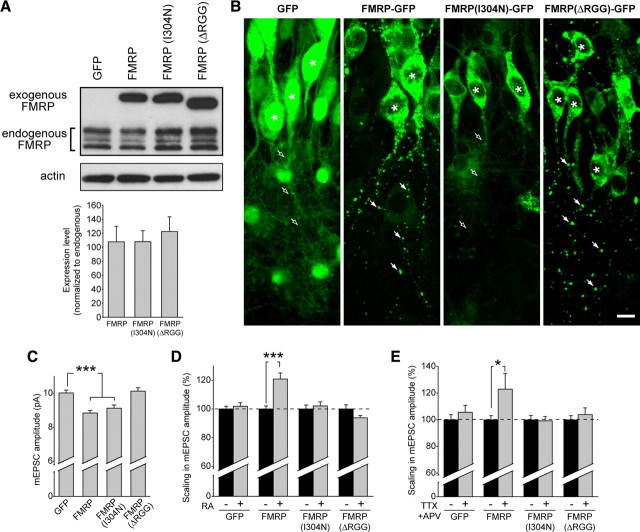
Viral expression of FMRP in knock-out slices restores synaptic scaling. A, Representative blot and quantification of exogenous FMRP–GFP, FMRP(I304N)–GFP, and FMRPΔRGG–GFP expression compared with endogenous FMRP protein levels after 6 d of virus expression in wild-type dissociated neurons (n = 6). B, Images from the CA1 region of hippocampal slices infected with lentiviral constructs expressing GFP, FMRP–GFP, FMRP(I304N)–GFP, or FMRPΔRGG–GFP. Asterisks indicate cell body. FMRP and FMRPΔRGG exhibit a punctate expression pattern in neuronal dendrites (filled arrows), whereas GFP or FMRP(I304N) are diffusely expressed in neuronal dendrites (open arrows). Scale bar, 10 μm. C, Amplitude of mEPSC events in KO neurons expressing GFP, FMRP, FMRP(I304N), or FMRPΔRGG (n = 38–66; ***p < 0.001). D, Percentage scaling after RA treatment in neurons expressing GFP or different FMRP constructs. DMSO groups for each construct were set to 100% to account for altered baseline amplitudes (see C) (n = 18–49; ***p < 0.001). E, Percentage scaling after treatment with 36 h of TTX + APV (n = 17–20; *p < 0.05). Error bars represent SEM.

