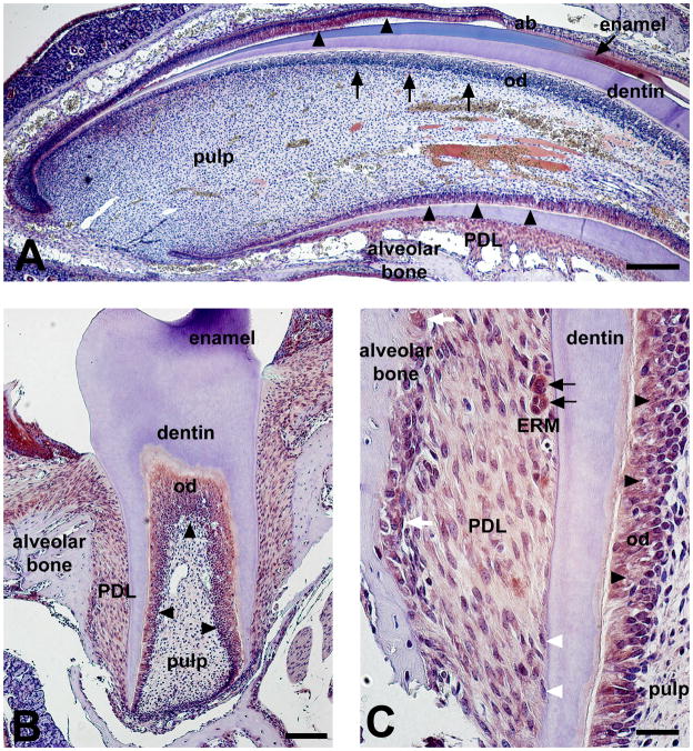
Figure 4.
Per2 expression in mouse incisor (A) and molar (B–C) at PN21 stage. Per2 showed distinct expression pattern at the root-analogue versus the root-analogue side in the incisor (A). Per2 was detected in root odontoblasts and ameloblasts (A, black arrowheads). In contrast, Per2 protein expression was low/undetectable in the odontoblasts of the crown-analogue side (A, black arrows). Dental pulp was devoid of staining in both incisors and molars (A–B). Odontoblasts in first molars were strongly stained for Per2 (B, black arrowheads). Per2 showed relatively weak expression in periodontal dental ligament (PDL) cells when compared with odontoblast expression (B–C). Epithelial rests of Malassez (ERM) showed strong expression of Per2 within the PDL space (C, black arrows). Per2 proteins are also detected in the nucleus of the osteoblasts and osteoclasts in the alveolar bone (C, white arrows). No Per2 protein expression is detected in cementoblasts (C, white arrowheads). ERM, epithelial rests of Malassez; od, odontoblasts; Scale bars = 200 μm in A, 100 μm in B, 20 μm in C.

