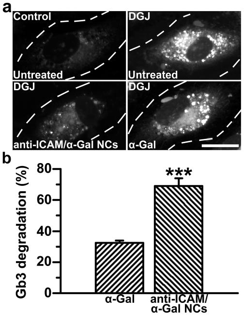
Figure 5. Anti-ICAM/α-Gal nanocarriers attenuate Gb3 accumulation in an endothelial cell model of Fabry disease.
(a) TNFα-activated HUVECs were incubated at 37°C for 16 h with fluorescent NBD-Gb3 and control cell medium or medium containing DGJ to inhibit endogenous αGal. Cells were then washed and left untreated or treated with α-Gal or non-fluorescent anti-ICAM/α-Gal NCs for 5 h, all in the presence of chloroquine to selectively permit activity of exogenous neutral α-Gal, and not endogenous acidic α-Gal. Cells were fixed and analyzed by fluorescence microscopy. Dashed lines mark cell borders, determined by phase-contrast. Scale bar, 10 μm. (b) Intracellular accumulation of NBD-Gb3 was quantified from micrographs as described in Materials and Methods. Data are mean±SEM (n≥72 cells). *** is p≤0.001, by Student’s t-test.

