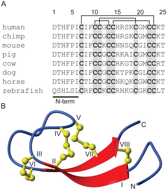
Figure 1.
Sequences and three-dimensional structure of hepcidin. (A). Sequence alignment of selected hepcidin sequences illustrating the high sequence conservation and the N-terminal region essential for biological activity. The sequences contain eight conserved cysteines that form four disulfide bonds, indicated by the lines above the sequence list. (B). A ribbon depiction of the three-dimensional structure of hepcidin showing the β-sheet structure (broad arrows) and the four disulfide bonds (ball and sticks).

