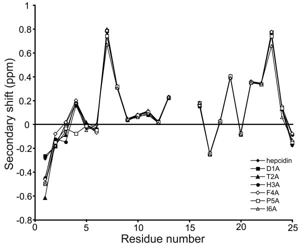
Figure 3.
NMR secondary shift analysis of the alanine mutants of hepcidin. All alanine mutants had almost identical αH secondary shift values to native hepcidin except for very small local changes around the site of substitution. A similar trend was observed for all the other hepcidin mutants analysed in this study (Figure S1).

