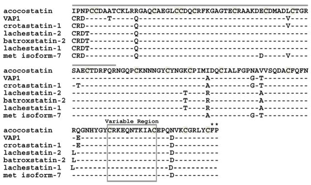
Fig. 6.
Amino acid comparison between acocostatin and six PIII disintegrin-like proteins. Dashes indicate amino acid identities as compared to the acocostatin sequence. The disintegrin-like domain is identified with a gray line above its sequence. The variable region is boxed. The first two amino acids of the hypervariable region are identified with asterisks (*). The variable and hypervariable regions are identified according to Takeda et al. (2006) VAP1 crystal structure and alignment analysis.

