Fig. 1.
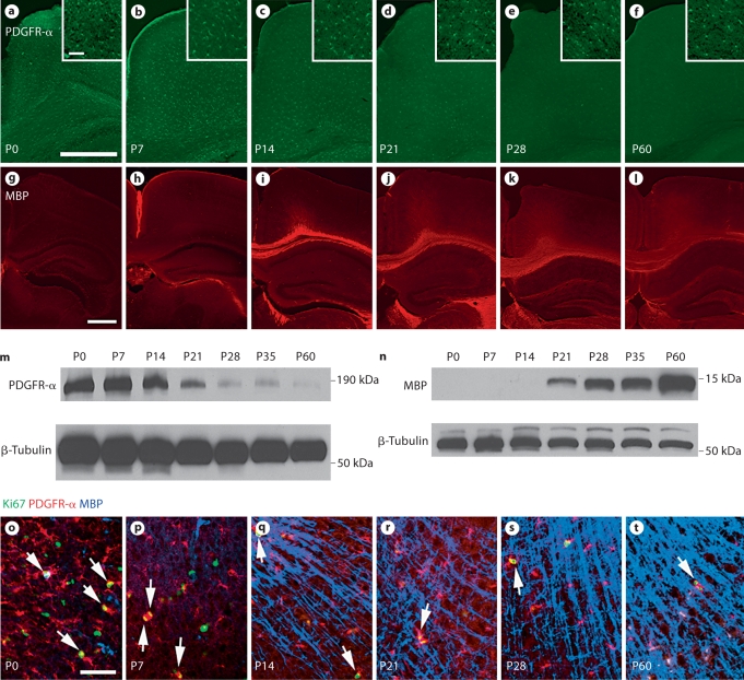
During normal development, the PDGFR-α-expressing oligodendrocyte progenitor population declines as the number of MBP-expressing mature oligodendrocytes increases. Cortical sections taken from wild-type animals at P0–P60 were stained for the oligodendrocyte progenitor marker PDGFR-α (a–f) and the mature oligodendrocyte marker MBP (g–l) using immunohistochemistry. Higher-magnification images of sections taken over the period from P0 to P60 stained with PDGFR-α, MBP and the proliferation marker Ki-67 demonstrate that proliferating oligodendrocyte progenitors diminish over time as myelination occurs, though proliferative PDGFR-α-expressing progenitors persist at least through 2 months of age (o–t, arrows). Scale bars = 500 μm (a, g) and 50 μm (a, inset, o). m, n Western blot analysis of cortical samples taken from animals ranging in age from P0 to P60 was used to characterize PDGFR-α (180 kDa; m) and MBP (14 kDa; n) expression.

