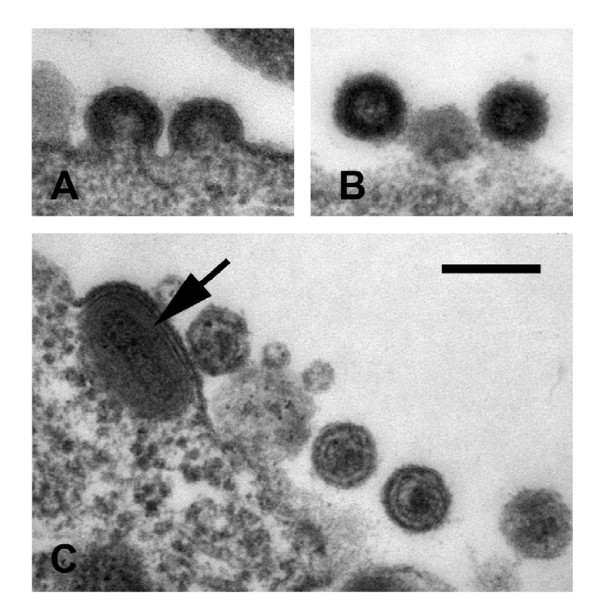
Figure 2.
Morphology of VLPs released from MVA-HERV-Kcon infected cells. (A) Virus assembly takes place at the cell membrane as is typically seen for C-type viruses. (B) Free immature particles and (C) mature particles with condensed cores could also be observed. Note the MVA particle (arrow) in the HERV-K-producing cell. Bar in (C) represents 200 nm.

