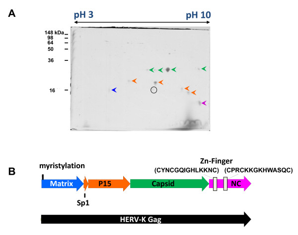
Figure 3.
Mapping of HERV-K Gag protease cleavage sites. (A) VLPs from MVA-HERV-Kcon-infected cells were pelleted by ultracentrifugation and separated by 2-D gel electrophoresis. The gel was stained with Coomassie and the indicated spots were analyzed by mass spectrometry and N-terminal sequencing. (B) Graphic scheme of HERV-K Gag, the experimentally determined processing products and in silico predictions. Colors indicate the analyzed spots isolated by 2-D gel electrophoresis.

