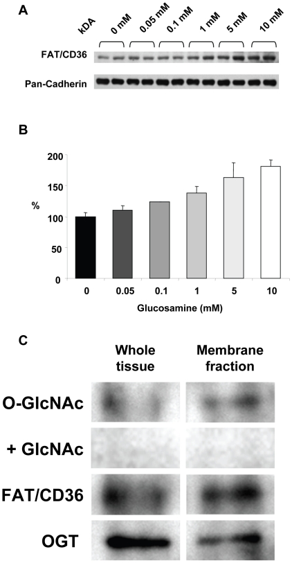
Figure 5. A) Immunoblots of Plasma membrane fraction for FAT/CD36 following 60 min perfusion with 0, 0.05, 0.1, 1, 5 and 10 mM; pan-cadherin included as a plasma membrane marker and protein loading control; B) Densitometric analysis of FAT/CD36 immunoblots normalized to 0 mM glucosamine; P<0.05 vs. 0 mM, one-way ANOVA with Dunnett's posthoc test; n = 2 in each group; C) Immunoprecipitation of FAT/CD36 from whole tissue and plasma membrane lysates, followed by O-GlcNAc and OGT immunoblots.
Specificity of O-GlcNAc antibody was confirmed by co-incubation with 10 mM N-acetylglucosamine (GlcNAc).

