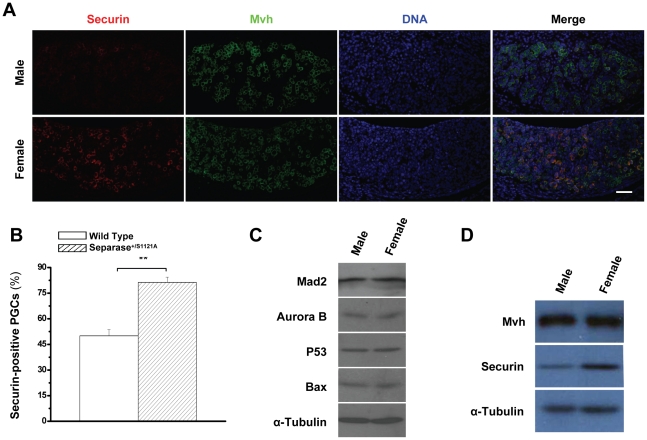
Figure 5. More Securin expression in female PGCs.
A. Securin levels of PGCs at 13.0 dpc observed by immuno-staining with Securin (red), Mvh (green) and DNA (blue). Bar = 5.0 µm. B. Quantitative analysis of Securin-positive PGCs in male and female gonads at 13.0 dpc. Serial sections of female genital ridges were stained for DNA, Mvh, and Securin. All Mvh-positive cells and the Mvh and Securin double positive cells were scored from at least three sections in at least three embryos (six genital ridges) for each time point. The mean value is shown with standard error (** p<0.001, T test). C. Western blot detection of Mad2, Aurora B, P53, and Bax of gonads at 13.0 dpc. D. Western blot detection of Mvh and Securin of gonads at 13.0 dpc.

