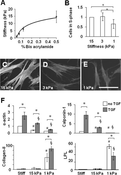
Figure 4.
Modulation of MSC proliferation and differentiation by matrix stiffness. (A) Stiffness of PA gels using 6% acrylamide and different bis-acrylamide concentrations. (B) MSCs were grown on PA gel for 1 day. Proliferation of MSCs on PA gels was quantified by BrdU incorporation. The number of cells in S phase for each sample was normalized to that on 15 kPa substrate to show fold-changes. * indicates significant difference (P<0.05). (C-E) Phalloidin staining of F-actin after 1-day culture. (F) MSCs were grown on collagen-coated stiff substrates or PA gels for 2 days in the absence or presence of TGF-β, and lysed for qPCR analysis. *, # and § indicate significant differences as in Figure 2A.

