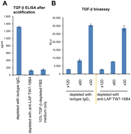
Figure 7. Immunologic depletion of “total” TGF-β from culture supernatants.
(A) A T cell culture supernatant from plate-bound anti-CD3/CD28 re-stimulated CD4+CD25− T cells cultured in 10% TGF-β-depleted FBS IMDM was treated with control IgG1 mAb-coated magnetic beads or anti-mouse LAP TW7-16B4 mAb-coated magnetic beads. The remaining “total” TGF-β in the culture supernatant was measured by TGF-β ELISA after acidification. Error bars represent mean ± S.D. of duplicates. (B) TGF-β activity in the same control IgG1-treated or anti-LAP TW7-16B4-treated T cell culture supernatant was measured by the 293T-caga-Luc-CD32-CD86 bioassay after ×30, ×60 and ×120 dilutions.

