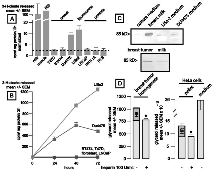
Figure 2.
Production of lipoprotein lipase activity by breast cancer, liposarcoma, and prostate cancer cells and in a breast cancer tissue sample. Panel A: Lipase activity is shown (mean ± SEM, 4 samples/group, corrected for cellular protein content and normalized to the value observed in milk (9 × 10³ cpm/2h). Human breast milk (50 μl), mouse gastrocnemius muscle (50 mcg protein, 45 × 103 cpm/2h), or tissue culture media conditioned by the indicated cell lines x 3 d were assessed for lipase activity (mean ± SEM, 4 samples/group, corrected for cellular protein content and activity observed in unconditioned media). The dotted line denotes the LPL activity found in unconditioned culture medium. Panel B: Time course of accumulation of lipase activity in conditioned culture media. Media (50 μl) were removed from cultures at the indicated intervals (mean cpm/mg protein ± SEM, n = 4 wells/timepoint). Panel C, upper: Identification of LPL in conditioned cell culture media. LPL was heparin-sepharose affinity purified from 10 ml fresh culture medium, 1.0 ml human breast milk, or 10 ml culture media conditioned (72 h) by LiSa-2 liposarcoma or DU4475 “triple negative” breast cancer cells, eluted with 0.6–0.8 M NaCl, and analyzed by western blot using anti-human LPL clone 43 (1:200). The band from milk was verified to contain LPL by mass spectrometry. Panel C, lower: Western analysis of a breast tumor homogenate (50 mcg protein without affinity purification) and breast milk (10 μl) for LPL. A band of the appropriate size is apparent in the tumor sample. Panel D: Estimation of the heparin-releasable LPL pool in breast cancer tissue and HeLa cells. Panel D left: tumor associated LPL activity is significantly reduced by heparin treatment (p = 0.0001). Panel D: right: Heparin reduced LPL activity residing in HeLa cell pellets by 29% (p < 0.04).

