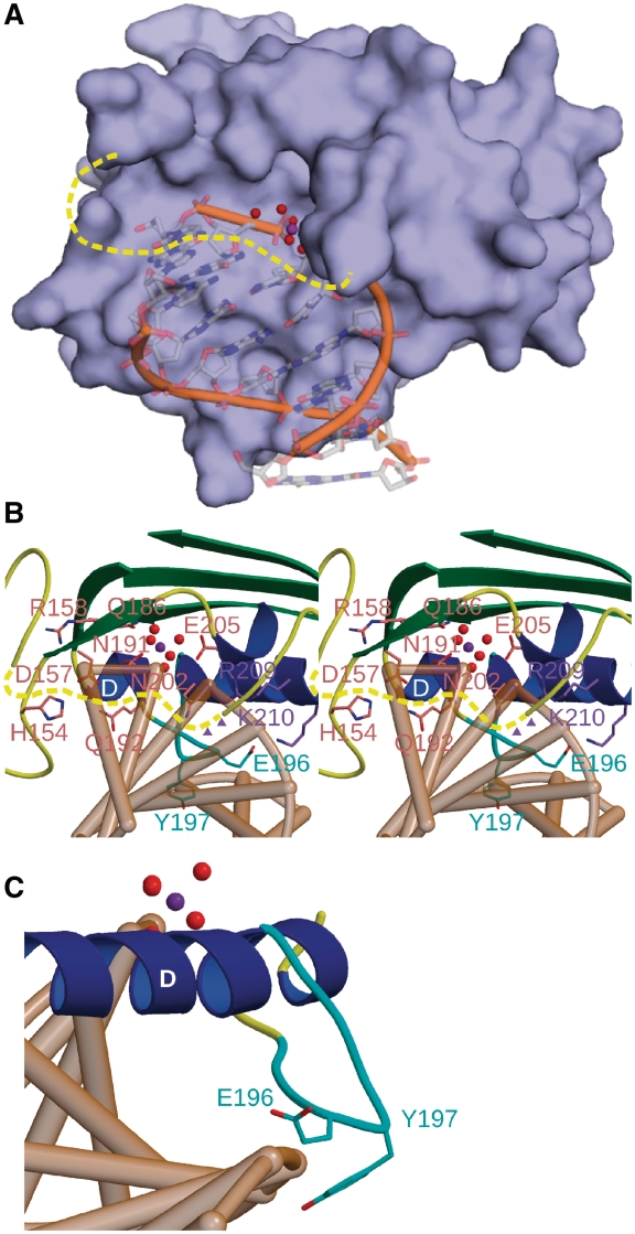
Figure 4.
Mutagenesis of catalytic and DNA-binding residues in the active site of EndA. (A) Surface rendering of EndA, with the modeled DNA substrate from the VVN nuclease [PDB ID code 1OUP (42)] superimposed in the active site. (B) Stereo ribbon diagram of the active site of EndA, displaying the ‘finger-loop’ (cyan), catalytic residues (pink) and DNA-binding residues (purple) targeted for mutagenesis. The disordered loop is drawn in dashed yellow. Putative positions of Arg127 and Lys128 on the disordered loop are shown as purple triangles. (C) Ribbon diagram of the ‘finger-loop’ position (cyan), relative to the putative position of a bound DNA substrate (tan).

