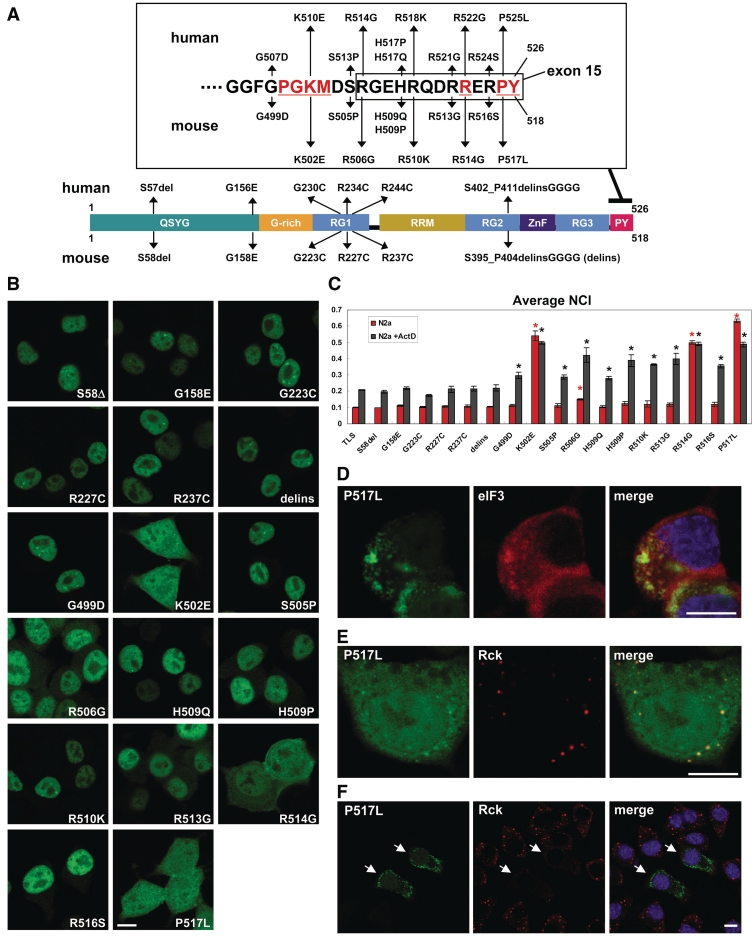
Figure 6.
Intracellular localization of ALS-linked mutants of EGFP-TLS. (A) ALS-linked mutations examined in this study. Mouse TLS mutations corresponding to known human mutations are indicated. Consensus residues of PY-NLS are shown in red and underlined. Residues encoded by exon 15 are boxed. (B) Intracellular localization of EGFP-TLS harboring above mutations in N2a cells. (C) Bars represent the average NCI (mean ± SD, n = 3) of ALS-linked mutants in untreated N2a cells (red bars) or actinomycin D-treated N2a cells (gray bars). *P < 0.05 in ANOVA and Dunnett's test in comparison with TLS. Red and dark asterisks indicate significant difference from TLS in the absence and presence of ActD, respectively. (D) An example of cytoplasmic granular structures formed by TLS mutants. (E) Partial colocalization of EGFP-P517L and PBs. EGFP-P517L showed partial colocalization with granules immunoreactive with anti-Rck. (F) Cells with higher expression of EGFP-P517L (arrows) showed a reduced number of PBs. PBs were immunostained by anti-Rck antibody. Scale bars represent 10 µm in (B, D, E and F).

