Figure 4.
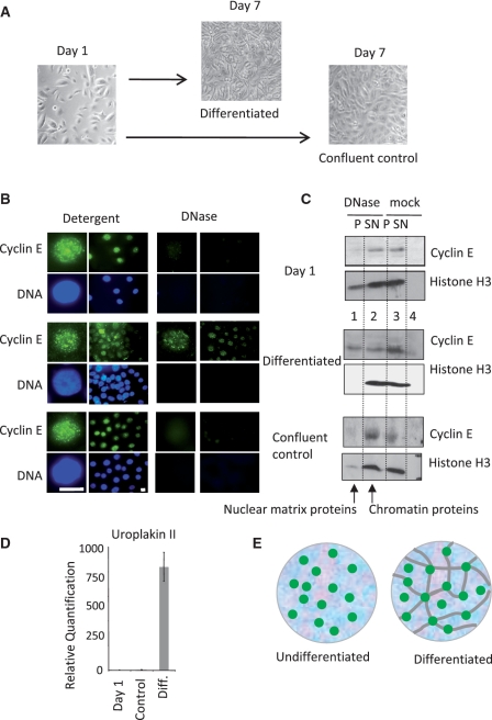
Recruitment of cyclin E to the nuclear matrix during differentiation of NHU cells (A) Cycling NHU cells were grown to confluence without differentiation or induced to differentiate over 7 days. (B) Cyclin E (green) and DNA (blue) in nuclei from cycling cells (Day 1, upper panels), differentiated (Day 7, middle panels) and confluent control cells (Day 7, lower panels), after extraction with detergent or DNase as indicated. Bar is 10 μm. (C) Detergent and DNase extracted protein fractions from the populations shown in A. Cyclin E changes from DNase sensitive in undifferentiated NHU and confluent control cells to partly DNase resistant in differentiated NHU cells, despite efficient extraction of histone H3. (D) Quantitative RT-PCR showing uroplakin 2 expression with SEM. (E) Model of cyclin E foci (green) associated with chromatin (blue background), unconstrained by attachment to the nuclear matrix (gray lines) in undifferentiated cells, but mounted upon a nuclear matrix in differentiated cells.

