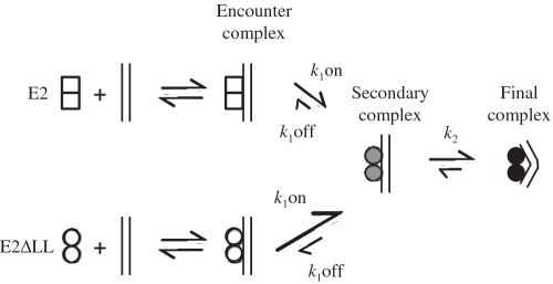
Figure 7.
A model for the E2-DNA complex formation. A schematic representation of the binding of 6 E2 and E2ΔLL to DNA. E2 dimers (open squares) bind to DNA to form a diffusion limited encounter complex. This subsequently forms a secondary complex (shaded circles) which in turn undergoes further structural rearrangement to form the final tightly bound complex (filled circles). We propose that the increased protein flexibility shown by E2ΔLL (open circles) facilitates formation of the secondary complex but has little or no effect on the following step. Formation of the secondary complex for E2ΔLL is then diffusion limited.

