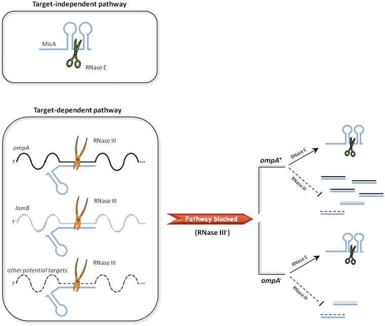
Figure 5.
Schematic representation of the two degradation pathways followed by MicA. RNase E and RNase III are represented by scissors and pliers, respectively. The two different pathways for MicA degradation are shown on the left side. The possible associations of MicA with its targets are also depicted. In the wild-type, MicA and the targets should be fully degraded as a result of both degradation pathways in cooperation with the exoribonucleolytic activity. In the RNase III− mutant, the MicA-target dependent degradation by RNase III is blocked. As a result, some degradation intermediates are stabilized and can be detected, namely the target and MicA strands that have interacted but could not be cleaved by RNase III. ompA being the main target of MicA, its species are over-represented. When additionally the ompA target mRNA is absent, the respective degradation intermediate is no longer present in the cell and, as a consequence, there is a reduced level of transcripts detected with probes complementary to MicA-targets.

