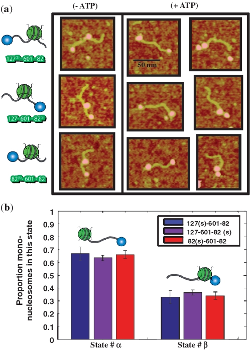
Figure 6.
RSC sliding of streptavidin labeled mono-nucleosomes. (a) Typical AFM images of streptavidin labeled mononucleosomes before and after the action of RSC and ATP for three different template constructions 127-601-82(s), 127(s)-601-82 and 82(s)-601-82 where (s) points for the end positioned streptavidin, and various naked DNA arm length (in bp) on each side of the 601 positioning sequence. (b) Proportion of streptavidin labeled mononucleosomes mobilized by RSC away from the streptavidin (State #α) or toward the streptavidin (State #β) for three different template constructions. The total number of streptavidin labeled mononucleosomes counted was N = 854 for 127-601-82(s), N = 551 for 127(s)-601-82 and N = 447 for 82(s)-601-82.

