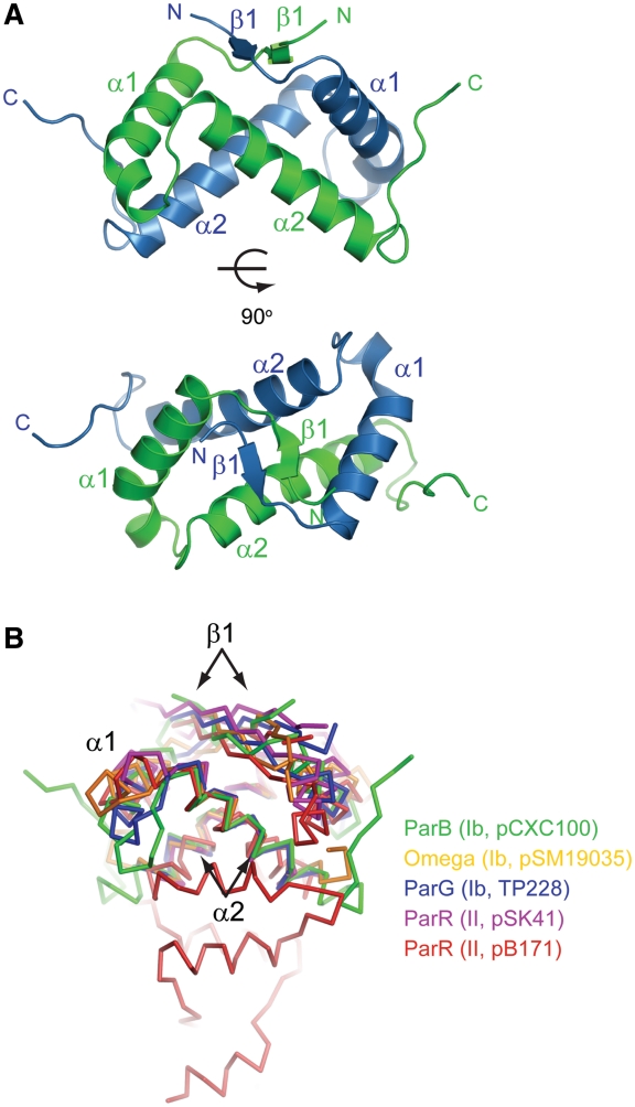
Figure 2.
Crystal structure of ParB DNA-binding domain. (A) Ribbon representation of the dimeric RHH structure in two orthogonal views. The two subunits are colored blue and green. Secondary-structure elements are labeled. The image on the bottom is viewed along the dyad axis of the dimer. (B) Structural superimposition of ParB (green) with other RHH CBPs: TP288 ParG (blue, 1P94), pSM19035 omega (orange, 2BNW), pSK41 ParR (magenta, 2Q2K) and pB171 ParR (red, 2JD3). These structures were aligned by their two α2 helices in PyMOL. The type and plasmid of the CBP are indicated.

