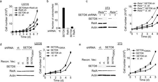
Figure 3. SETD6 attenuates RelA-driven cell proliferation.
(a) Increased proliferation rates of SETD6-depleted U2OS cells required RelA. Cell number in the indicated lines was determined daily for seven days. (b) Enhanced anchorage-independent growth of SETD6-depleted cells required RelA. Quantitation of colonies per field in soft agar assays with the indicated cell lines. (c) Increased proliferation rate of SETD6-depleted MEFs required RelA protein. Rela+/+ and Rela−/− MEFs stably transduced with SETD6 shRNA or control shRNA and assayed as in (a). Left panel: immunoblot analysis of WCE from the indicated cell lines. (d–e) Catalytic activity of SETD6 was required to restore a normal growth rate to SETD6-depleted cells. Growth curves and WCE immunoblot analysis of U2OS (d) and 3T3 fibroblast (e) cells reconstituted with SETD6 or SETD6Y285A as indicated. Error bars in (a–e) indicate ± s.e.m. from at least three independent experiments.

