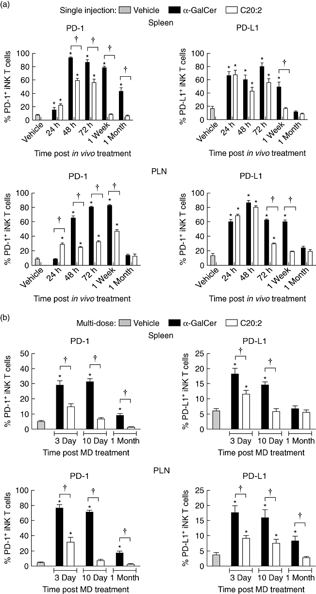
Fig. 4.

Kinetic analysis of up-regulation of programmed death-1 (PD-1) and programmed death ligand-1 (PD-L1) surface expression on invariant natural killer T (iNK T) cells following glycolipid administration. (a) Non-obese diabetic (NOD) mice (4–6 weeks old) were injected with a single dose [4 µg, intraperitoneally (i.p.)] of glycolipid or vehicle and rested for various times (24 h, 48 h, 72 h, 1 week and 1 month). Spleen and pancreatic lymph node (PLN) lymphocytes were isolated, and the levels of PD-1 and PD-L1 surface expression on T cell receptor (TCR)-β+tetramer+ iNK T cells were analysed by flow cytometry. (b) NOD mice (4–6 weeks old) were treated with a multi-dose (MD) protocol of α-galactosylceramide C26:0 (α-GalCer), C20:2 or vehicle (4 µg/dose every other day for 3 weeks, i.p.) and rested for either 10 days or 1 month. Spleen and PLN lymphocytes were isolated and the levels of PD-1 and PD-L1 surface expression on TCR-β+tetramer+ iNKT cells were analysed by flow cytometry. Data are representative of two independent experiments yielding similar results and are expressed as mean ± standard deviation (s.d.); five mice/treatment group/time-point. *Significant (P < 0·05) difference between the vehicle and glycolipid treatment groups. †Significant (P < 0·05) difference between α-GalCer and C20:2 treatment values.
