
Fig. 1.
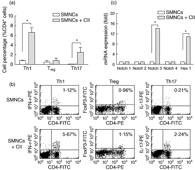
Collagen-specific reactivation tends to T helper type 1 (Th1)- and Th17-type expansion along with activated Notch signalling and increased Notch3 expression. (a) Spleen mononuclear cells (SMNCs) from collagen II (CII)-immunized DBA/1J mice were cultured in vitro with or without CII; 3 days later, cells were collected and the percentage of Th1, regulatory T cells (Treg) and Th17 cells were analysed using flow cytometric intracellular staining, as described in Methods. (b) The representative flow cytometric results summarized in (a) are shown; the percentages of relative cytokine- or transcript factor-expression T cells are indicated in the dot-plots. (c) After 3 days' culture with or without CII, CD4+ T cells were purified from SMNCs by magnetic sorting kits and were assessed for transcript levels of Hes1 and four Notch receptors, including Notch1, Notch2, Notch3 and Notch4 by real-time polymerase chain reaction (PCR). *P < 0·05.