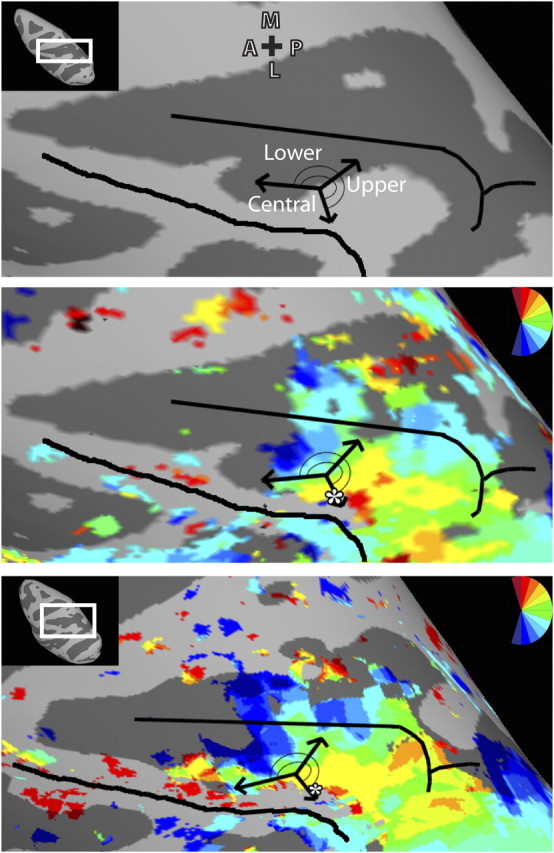
Figure 8.

Comparison of LIP topography based on physiology and fMRI data. Top, Schema of LIP topography adapted with permission from Ben Hamed et al. (2001) (their Fig. 8C). The fundus of the IPS and crown of the inferior parietal lobule are denoted by black lines. A representation of the upper visual field was found in posterior LIP, and the lower visual field representation was found anterior. A representation of central space was identified on the lateral border of LIP. Middle and bottom, The schema of LIP topography defined by physiology data overlaid upon the topography of LIPvt from the LH of monkey M1 (middle) and the group map (bottom) revealed by fMRI. There is strong correspondence of LIP topography between the physiology and fMRI data with the upper visual field progressing anterior and medial to a lower visual field representation, and a representation of central space on the lateral border (white asterisk).
