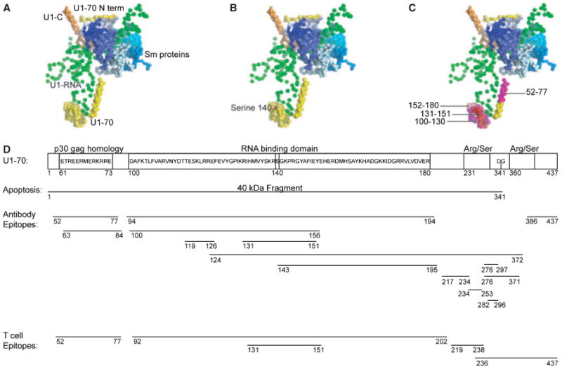
Fig. 2. Modifications and autoimmune epitopes on U1-70K.

(A) The U1-snRNP crystal structure (PDB ID: 3CW1) (7) was visualized using Pymol. In this depiction of the structure, U1-RNA is depicted in green, U1-70K in yellow, U1-C in orange, and the Sm proteins are displayed in blues. U1-A is not included in this depiction, as it was not part of the crystal complex in this structure. (B) The phosphorylation of serine residue 140 is a modification that occurs during apoptosis, and is shown in magenta. (C) The domain (100–180) of U1-70K is highlighted and is the RNA-binding domain of the protein. Notably the B-cell and T-cell epitope 131–151 is shown in red. The residues around this epitope are highlighted: 100–130 in magenta, and 152–180 in light pink. These regions contain additional B-cell and T-cell epitopes. (D) The domains of U1-70K include an N-terminal domain, RNA-binding domain, and serine/arginine repeats. During apoptosis, a 40 kDa fragment is cleaved at residue 341. B-cell and T-cell epitopes on U1-70K are shown.
