
Figure 2.
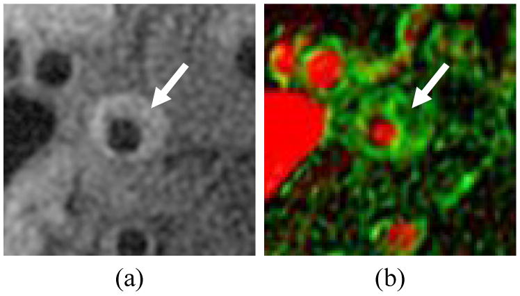
Vasa vasorum image of a carotid artery showing a) corresponding T1-weighted image, b) vasa vasorum image with vp mmapped to the red channel (full red corresponds to100%) and Ktrans mapped to the green channel (full green corresponds to 0.2 min-1). Arrow indicates eccentric plaque with high Ktrans.