
Figure 3.
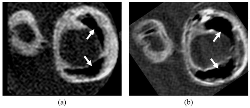
Matched cross sections of a carotid endarterectomy specimen imaged on a) 3 Tesla and b) 7 Tesla whole body scanners. At 7 Tesla, the specimen exhibits an apparent increase in the size of calcifications (arrows), more conspicuous variations in wall contrast, and overall higher signal-to-noise ratio.