
Figure 4.
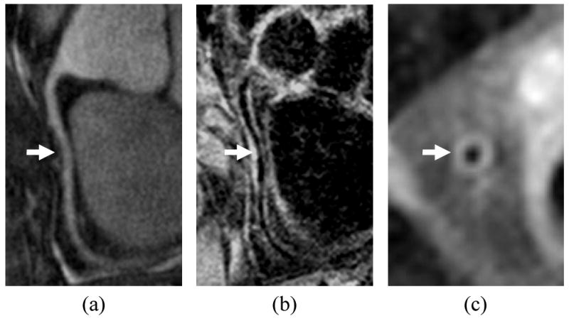
Coronary wall imaging showing a) MRA of right coronary artery, b) longitudinal vessel wall image with thickening indicated by arrow, c) cross sectional vessel wall image. Blood suppression was accomplished using motion-sensitized driven equilibrium, which does not depend on inflow over the long course of the artery.