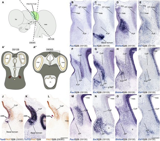
Figure 8.
Dorsoventral extent of seven pretectal markers in the quail, studied by in situ hybridization/immunoreaction in transversal cryostat sections at Q26 (two specimens). (A,A′,A′) Schemata showing a lateral view with two variant transversal section planes used (see specimen code in brackets) and the respective aspect of such transversal sections, in order to illustrate the brain region analyzed in both specimens. [Block comprising (B–I,M–P)] comparisons of four markers (Pax3, Six3, Bhlhb4, Ebf1) at three section levels from rostral to caudal from a single specimen (levels arranged top to bottom along the columns). (J–L) Three adjacent transversal sections at CoP level, allowing comparison of Pax6, PAX7, and Tcf7l2 dorsoventral patterns in a different quail specimen. The red arrowhead in (L) shows the ventral-most limit of PAX7 expression, slightly above the alar–basal boundary(ABB; black arrow). Numbers between brackets: code of the specimen in our collection. Scale bar = 300 μm.

