Fig. 1.
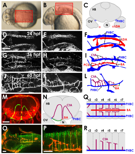
Vascular anatomy of the developing zebrafish hindbrain. (A,B) Transmitted light images of 30 hpf zebrafish, with boxes indicating the dorsal (A) and lateral (B) regions of the hindbrain imaged here. (C) Schematic transverse section of the hindbrain of a 24 hpf zebrafish, showing the locations of the paired venous primordial hindbrain channels (PHBCs) relative to the hindbrain, otic vesicles, notochord and pharyngeal arterial lateral dorsal aortae (LDA). N, notochord; HB, hindbrain; OV, otic vesicle. (D-L) Confocal images of the hindbrain vasculature in Tg(fli1a:EGFP)y1 (D,E,G,H) or Tg(kdrl:GFP) (J,K) embryos at 24 (D-F), 36 (G-I) or 60 (J-L) hpf, with explanatory schematic diagrams of the vasculature at each stage (F,I,L). Images and diagrams include dorsal (D,G,J), dorsal-lateral (E,F,H,I) or lateral (K,L) views. Rostral is to the left. Diagrams show LDA (red), PHBC (blue), basilar artery (BA, red) and central arteries (CtAs, purple). (M) Confocal image of a transverse section at the hindbrain level through a 3 dpf Tg(kdrl:GFP) embryo (green, blood vessels), counterstained with Rhodamine-phalloidin (red, F-actin). (N) Explanatory diagram of the blood vessels and major anatomical structures in M. Connections (green) persist between the PHBC and BA underneath the hindbrain. (O,P) Whole-mount immunofluorescent staining of blood vessels with anti-GFP (red) and of paraboundary cells with anti-DeltaD (green) in a 60 hpf Tg(kdrl:GFP) transgenic embryo. Lateral view, rostral to the left. The boxed area is shown at higher magnification in P. The anti-DeltaD staining efficiently labels rhombomere paraboundary cells, permitting visualization of rhombomere boundaries (dashed lines). (Q,R) Dorsal (Q) and lateral (R) diagrams summarizing the pattern of growth, interconnection and BA linkage of the CtAs. r, rhombomere. Scale bars: 50 μm.

