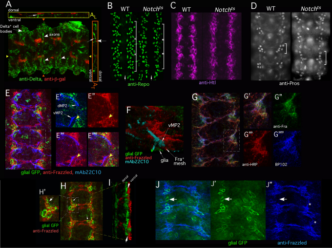
Fig. 2.
Interface glia recruit a cap of Frazzled+ neuronal processes. The CNS of stage 13 Drosophila embryos was analyzed by immunofluorescence. Embryos in E-G and J are very early stage 13, other panels are mid-stage 13. (A) Stage 13 embryos bearing E(spl)mδ-lacZ reporter, labeled with anti-β-gal (red) and anti-Delta (green). A z-projection is shown in A. White arrowheads indicate Delta-positive axons coursing medially from lateral neurons. Anterior is to the right. AL shows a longitudinal cross-section at the position of the yellow triangles. AT shows a transverse cross-section at the position of the orange triangles. Thin white arrows indicate Delta-positive axons (green) as they contact Notch signaling-positive glia (red). (B) Wild-type (WT) or Notchts embryos were temperature-shifted, fixed and labeled with anti-Repo. Each white bracket indicates one segment. White arrow indicates midline. Wild-type and Notchts embryos each have approximately ten Repo+ interface glia per hemisegment. A few non-interface glia are also visible in this section. (C) Wild-type or Notchts embryos prepared as described for B but visualized with anti-Htl. (D) Wild-type or Notchts embryos prepared as described for B but visualized with anti-Prospero. Five to six Pros+ cells are visible in each segment in wild type and in Notchts (bracket). Only one focal plane is shown so not all Pros+ cells are visible in all segments. (E-E″″) Wild-type embryo expressing mCD8-GFP in interface glia, under the control of htl-GAL4, labeled with anti-GFP (green), anti-Frazzled (red) and mAb 22C10 (blue). E shows the overlay of all channels. E′-E″″ are higher magnification views of the boxed region. Thin white arrows indicate the growth cones of dMP2 and vMP2. Glia (GFP, green) and growth cones (mAb 22C10, blue) are shown in E′. Anti-Frazzled (red) is shown in E″. Anti-Frazzled (red) and growth cones (mAb 22C10, blue) are shown in E‴. E″″ is the overlay of all markers. Yellow arrows highlight the gap between the vMP2 growth cone and the glial cell. Note close apposition of dMP2 and vMP2 growth cones with Frazzled. (F) Three-dimensional rendering of an image stack of an embryo prepared as described for E. vMP2 growth cone (mAb 22C10, cyan) migrates through a bed of Frazzled+ processes (red) that separate it from the associated interface glia (GFP; green). Image has been inverted to put glia beneath axon. (G-G″″) Wild-type embryo labeled with anti-Frazzled (green), anti-HRP (red) and mAb BP102 (blue). G shows overlay of all channels. A single optical section is shown (~0.7 μm). G′-G″″ show higher magnification views of the boxed region. G′ shows the overlay of channels; G″-G″″ show separate channels as indicated. (H-I) An embryo expressing mCD8-GFP in interface glia (htl-GAL4), labeled with anti-GFP (green) and anti-Frazzled (red). H is a z-projection. Arrows highlight the correspondence of glial position and Frazzled. H′ is a magnified view of the region indicated in H. I shows a three-dimensional rendering of H, viewed obliquely. Frazzled is ventral to the glia (arrows). (J-J″) z-projection of an early stage 13 wild-type embryo expressing mCD8-GFP in interface glia (GAL4-605), labeled with anti-GFP (green) and anti-Frazzled (blue). Frazzled+ cap is beginning to fill-in between successive segments (asterisks). J shows the overlay of channels. Arrows indicate an intersegmental region in which Frazzled is not yet detected but a glial cell is already present. J′ shows GFP signal in glia and J″ shows anti-Frazzled staining. At early stage 13 GAL4-605 is expressed primarily in interface glia; later it will also be in some neurons.

