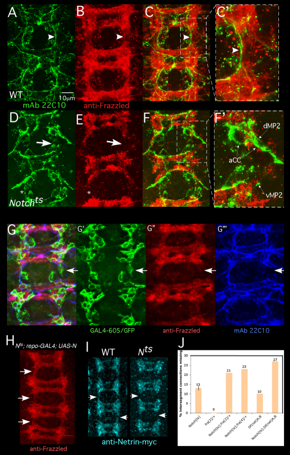
Fig. 3.
Notch is required for recruitment of a Frazzled- and Netrin-rich neuronal cap by interface glia. (A-F) Drosophila embryos were prepared as described in Fig. 1 legend and stained with mAb22C10 (green) and anti-Frazzled (red). A wild-type embryo is shown in A-C′. Boxed region in C is shown in C′. Arrowheads highlight correspondence between axon trajectory and Frazzled distribution. A Notchts is shown in D-F′ Boxed region in F is shown in F′. Thick arrows highlight a gap in the Frazzled pattern and a corresponding gap in the longitudinal tract. Thin arrows indicate stalled dMP2 and vMP2 growth cones. In some segments, axons cross between neuromeres despite a gap in Fra accumulation (asterisk). (G-G‴) Notchts embryos expressing mCD8-GFP in interface glia (GAL4-605) were fixed and labeled with anti-GFP (green), anti-Frazzled (red) and mAb 22C10 (blue). Overlay of all channels is shown in G. White arrows indicate an intersegmental connection that has failed to develop. G′-G‴ show separated channels, documenting a gap in the Frazzled pattern and a corresponding break in the early axon scaffold despite the presence of an interface glial cell in the gap. (H) A Notchts; repo-GAL4; UAS-Notch embryo was prepared as described for A-G and visualized with anti-Frazzled. Expression of Notch in glia rescues Frazzled distribution in nearby neurons (arrows; compare with B,E,G″). (I) Wild-type or Notchts embryos bearing a chromosomal NetrinB-myc ‘knock-in’ (Brankatschk and Dickson, 2006) were temperature-shifted, fixed and visualized with anti-myc. Netrin-myc spreads between segments in wild type, but gaps persist in Notchts (Nts; arrows). (J) Flies of the indicated genotype were temperature-shifted and intersegment connections were assayed with mAb22C10. The thin vertical line on the Notchts bar shows s.e.m.

