Fig. 4.
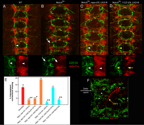
Different mechanisms for glial versus neuronal rescue of longitudinal pioneers by Notch. (A-D) Early stage 13 wild type (A), Notchts (B), Notchts; repo-GAL4; UAS-Notch (C; glial rescue) and Notchts;15J2-GAL4; UAS-Notch (D; pioneer-specific rescue) embryos were visualized with mAb 22C10 (green) and anti-Frazzled (red). Indicated regions in each panel are shown in insets beneath. Arrowheads in A and C highlight correspondence between Frazzled and pioneer axon trajectory; arrows in B indicates gap in Frazzled and corresponding break in longitudinal track in Notchts; yellow arrow and arrowheads in D indicate discordance between rescued axon growth but residual broken Frazzled pattern upon restoration of Notch to pioneers. (E) Embryos were prepared and scored as for the experiments shown in Figs 1, 2 and 3; bars indicate percentage of intersegment connections missing at mid-stage 13. Orange bars indicate rescue by expression of transgenes in glia; cyan bars indicate rescue by expression in dMP2 and vMP2. Statistical significance relative to Notchts is shown by asterisks: *P<0.05; **P<0.01 (ANOVA). (F) Wild-type embryo expressing mCD8-GFP in dMP2 visualized with anti-Delta (green) and anti-GFP (red). Yellow arrow indicates dMP2 growth cone as it contacts Delta+ commissural axons (white arrows). dMP2 cell body is marked with an asterisk.

