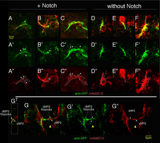
Fig. 6.
Notch promotes presence of filopodial structures in dMP2. (A-F″) Notchts; 15J2-GAL4 embryos expressing actin-GFP in dMP2 with (A-C″) or without (D-F″) wild-type Notch were temperature-shifted and fixed to preserve the actin cytoskeleton and visualized with anti-GFP (green) and a marker for the microtubule cytoskeleton, Futsch (mAb22C10, red). A gallery of single growth cones is shown. Middle row shows actin-GFP signal; arrows highlight whisker-like projections that label for actin-GFP but not Futsch. [Projections that co-labeled with Futsch (D,F) were interpreted as branches rather than filopodia and were not counted.] Bottom row shows Futsch signal with outline of actin signal superimposed. At this stage, 15J2-GAL4 expression is stronger in dMP2 than vMP2, allowing us to visualize this pioneer alone. Futsch expression in nearby cells accounts for unassigned red label. Mean length of single filopodia (2.2±0.1 μm) and maximum length (4.5 μm), were not significantly different with and without Notch, and were similar to dimensions observed in live imaging (Homem and Peifer, 2009). (G-G″) A GFP-labeled dMP2 growth cone (green, left) encounters the converging vMP2 (right) in an embryo prepared as described for A-F. Note the dMP2 filopodia embracing the oncoming vMP2 (G′). The prominent 22C10+ growth cone is vMP2; the central domain of the dMP2 growth cone is also indicated in mAb22C10 channel (G″; dashed arrow). GT shows a cross-section through the vMP2 growth cone (red) and dMP2 filopodia (green), at the position of the yellow triangle. Anterior is to the left.

