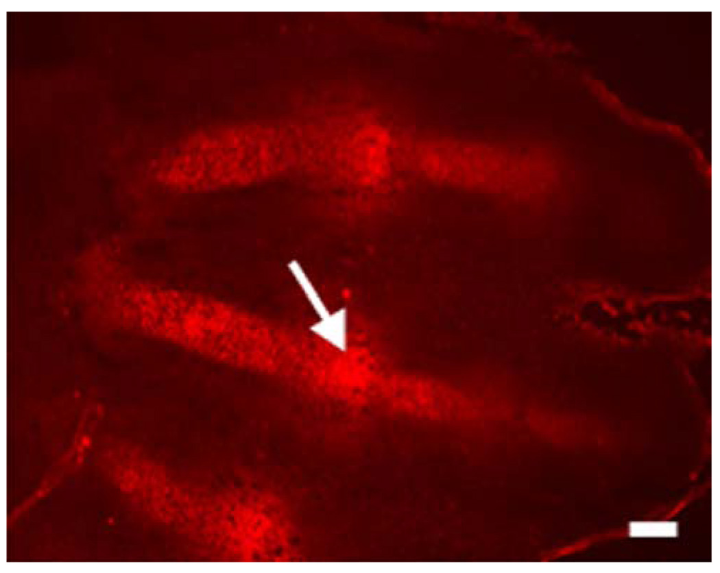
Fig. 3.
Histological section of the digital rays of an autopod of a mouse at embyonic day 13.5 (E13.5). Staining with the marker of hypoxia, EF5 (red), shows that the chondrocytes are hypoxic in the digital rays. The “interzones”, which will give rise to the perspective joints, are also highly hypoxic (white arrow). Bar 100 µm

