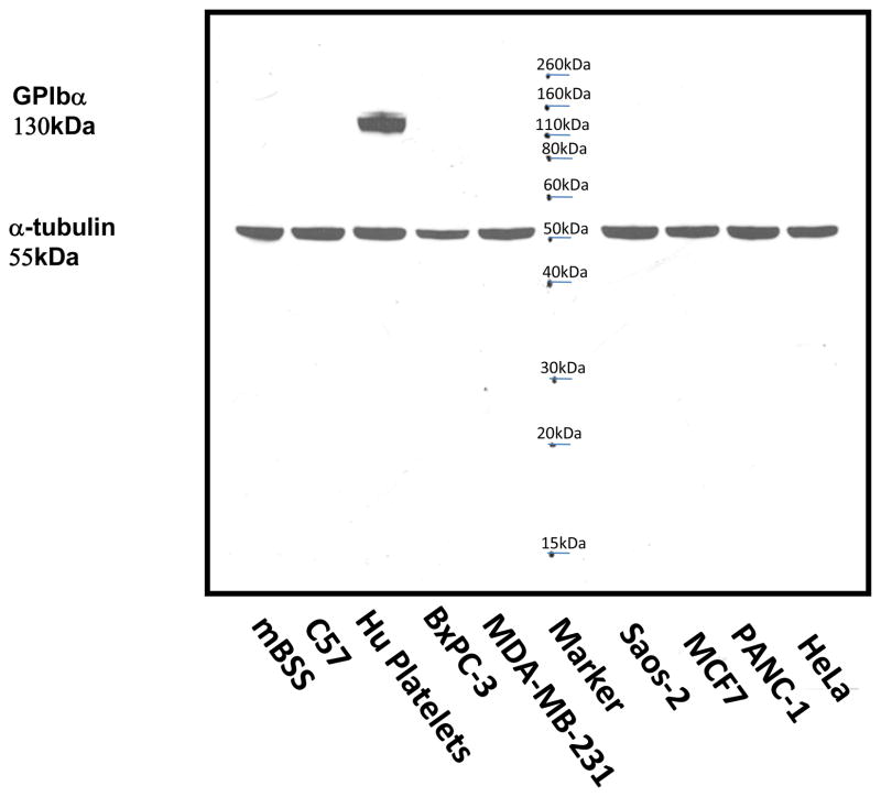
Figure 3.
Western blot analysis of human tumor cell lines for the presence of human GP Ibα antigen. Also shown are normal mouse and human platelet lysates for representative signals. The blot was reacted with an anti-α-tubulin antibody as a positive control. Human GP Ibα antigen is observed in lysates from purified human platelets (Hu Platelets) and noticeably absent in all other samples (anti-human GPIbα monoclonal antibody, LJ-Ib10, kindly provided by Zaverio Ruggeri, The Scripps Research Institute).mBSS, mouse platelet lysate missing GPIbα; C57, normal mouse platelet lysate; Hu Platelets, human platelet lysate. All other lanes contain lysate from the indicated human tumor cell line.

