Fig. 6.
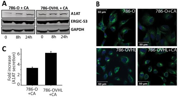
CA modulates ERGIC-53 activity but not its levels. (A) Intracellular levels of ERGIC-53 and its client A1AT were monitored over 24 h after treatment with 0.01 μmol/L CA via western blotting. (B) Effect of CA on distribution of ERGIC-53 in 786-O and 786-OVHL cells visualized by immunofluorescence microscopy. Cells were treated with either DMSO (control) or 0.01 μmol/L CA for 24 h. Note the diffuse staining for ERGIC-53 (green fluorescence) in the perinuclear area in CA treated 786-O cells. (C) A1AT levels in conditioned medium obtained from 786-O and 786-OVHL cells treated with 0.01 μmol/L CA were measured 24 h after treatment using ELISA. Values were then normalized to cell number and are reported relative to controls. Data represent the average of three experiments ± SEM.

