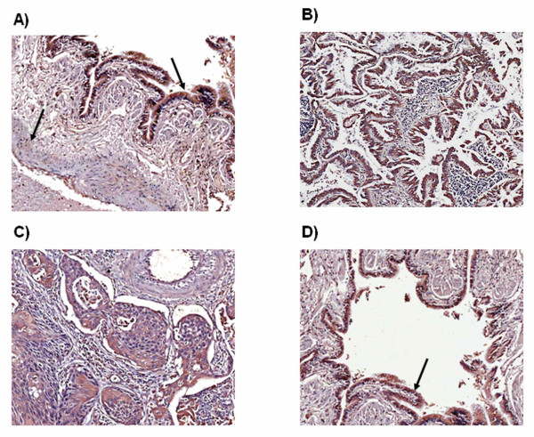
Figure 2.
Expression of PGIS and TXS in a retrospective panel of tumour/normal matched tissue samples. TXS was weakly expressed in the smooth muscle of normal pulmonary vessels, with a moderate expression observed in pulmonary epithelial cells (A). In tumour sections, TXS expression was observed to a varying degree in both adenocarcinoma (B) and squamous cell carcinoma (C) tissue. A weak TXS expression was observed in vascular smooth muscle cells of the tumour vasculature (C), with strong expression observed in tumour epithelial cells (D). Magnification ×10.

